Expert Sonography Services for Early Detection

Experienced sonologists specializing in advanced imaging techniques for optimal health outcomes.

Advanced Imaging Solutions

Doppler, 3D/4D, and musculoskeletal imaging expertise.

Early detection leads to timely and effective treatment.

Trust our expertise for your imaging needs.

Doppler Imaging
3D/4D Imaging

About Us

Experienced sonologists dedicated to early detection and effective treatment through advanced imaging techniques.

An ultrasound machine displaying an image on its screen is situated on a hospital trolley. The machine has a covered keyboard and several wires or cables attached. The setting appears to be a medical environment, likely a hospital or clinic, with a sterile and clinical appearance.
A person wearing blue gloves is operating an ultrasound machine. The control panel is visible with various buttons and dials, and two screens display information. There is also a small bottle in a holder next to the machine.

Expert Imaging Services

Providing advanced sonography with experienced sonologists for early detection and effective treatment solutions.

Doppler Imaging

Specialized in Doppler imaging for accurate assessment of blood flow and vascular conditions.
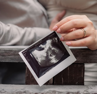
A printed ultrasound scan placed on a shiny, crumpled surface that reflects light. The scan shows a developing fetus within the womb, displaying typical ultrasound colors in shades of black, white, and gray.
3D/4D Imaging

Offering 3D/4D imaging services for detailed visualization of anatomical structures and fetal development.

Expert musculoskeletal imaging for precise diagnosis and treatment planning of joint and soft tissue conditions.

Musculoskeletal Imaging
A person wearing blue medical scrubs and gloves operates an ultrasound machine in a clinical setting. The individual's hands are adjusting the controls and handling the transducer probe, which is resting on the console equipped with various buttons and dials.
A medical imaging device with a monitor displaying a user interface featuring multiple icons and buttons. The device is labeled 3D OCT Maestro by Topcon. There is also a black adjustable arm and other medical supplies in the background.

Exceptional care and expertise in sonography. Early detection made all the difference for my health.

A. Priya

A medical professional is performing a procedure on a patient's leg in a clinical setting. The patient is lying on a table with blue surgical drapes covering most of their body. An ultrasound machine displays an image, possibly of the patient's leg, while a monitor hangs on the wall behind them.
A close-up of hands holding an ultrasound image against a wooden background. The focus is on the photograph of the ultrasound, which is being gently grasped by a hand with manicured nails, showing a ring on one finger. The background and clothing suggest a casual setting.

★★★★★

Gallery

Explore our advanced imaging techniques and expert sonologists' work.

Two people holding a printed ultrasound image together, each using one hand. The ultrasound image is black and white, indicating a medical scan, possibly of a fetus. The individuals are wearing light-colored clothing, and one of them has a ring on their finger.
A newborn baby is being examined by medical professionals. One person is using an ultrasound device while another uses a stethoscope to check the baby's health. The baby lies on a white cloth, surrounded by individuals wearing gloves, emphasizing a clinical setting.
A stethoscope and a sphygmomanometer are laid out on a white fabric surface. The stethoscope has a shiny metallic diaphragm and black tubing. The sphygmomanometer features a round gauge with blue, red, and black markings indicating blood pressure measurements, attached to a gray and black bulb used for inflating the cuff.
A black and white image of a couple holding sonogram pictures. The woman, wearing a floral dress and bracelets, lovingly gazes at her partner, who smiles back. The focus is on the sonogram images being held by the couple, capturing a sense of joy and anticipation.
A person wearing a white shirt and blue jeans is holding several ultrasound images. The focus is on the hand and the ultrasound pictures, suggesting an intimate or meaningful context. Sunlight casts soft shadows on a wooden floor.